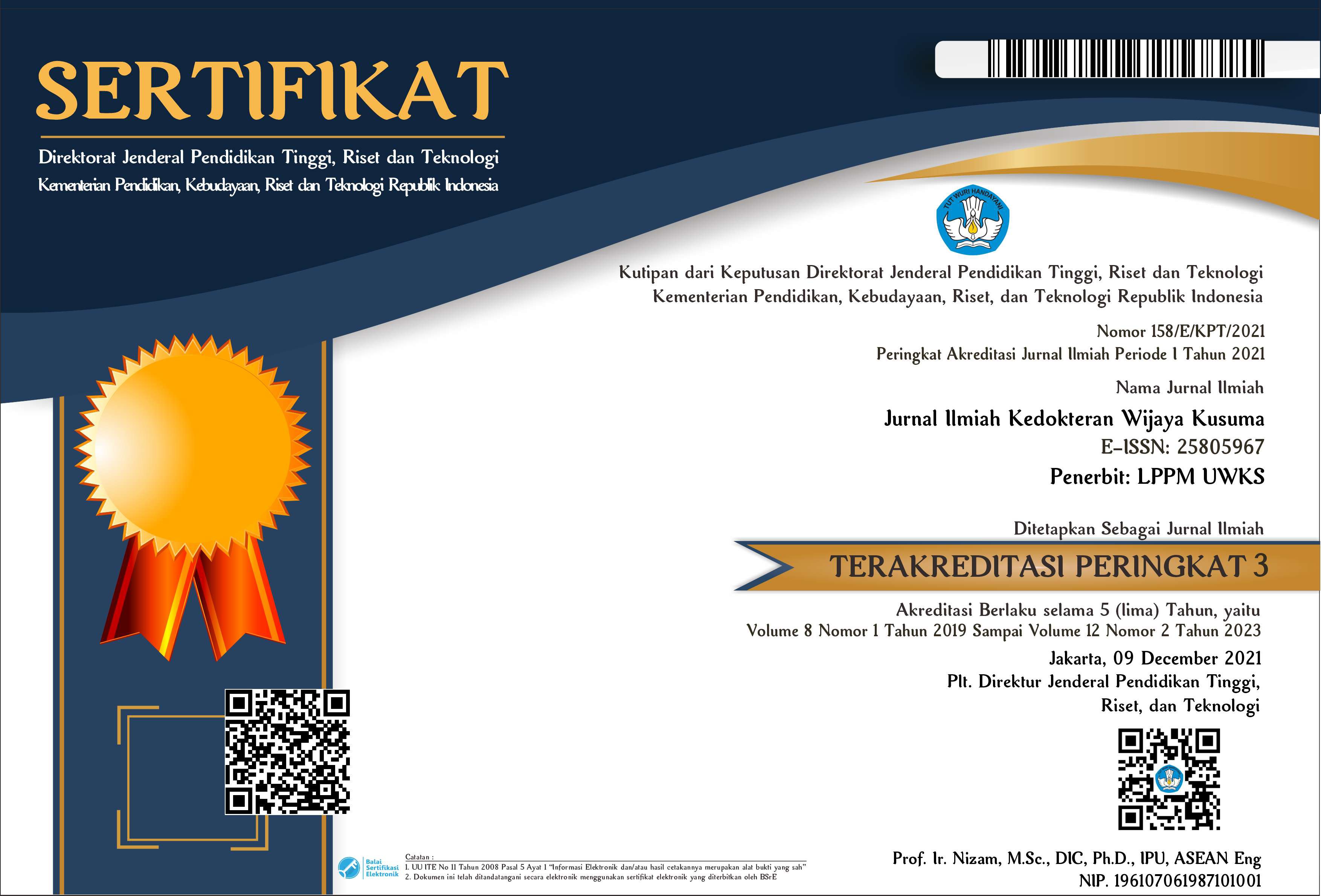
Increased Levels of Nerve Growth Factor Indicating Brain Injury In Mice Model
DOI:
https://doi.org/10.30742/jikw.v11i2.2024Keywords:
nerve growth factor, hypoxic ischemic, Wistar ratsAbstract
Hypoxic-ischemic encephalopathy (HIE) brain injury is one of the leading causes of death and disability worldwide. Nerve growth factor (NGF) is a neurotrophin that plays an important role in the natural repair and regeneration of nerves, but the previous study regarding NGF level after brain injury is still scant. This study aims to determine NGF levels in male Wistar rat models that received right Common Carotid Artery (CCA) occlusion. This study used an experimental and control design conducted in July-August 2021 at the Stem Cell Research and Development Center, Universitas Airlangga. The right CCA occlusion was performed on the Wistar mouse model in the treatment group, then placed in a hypoxic chamber and reperfusion after 60 minutes. Observations of neurology scores were carried out in the first 24 hours. After 2x24 hours the animal was sacrificed for serum NGF level measurement using the ELISA method. Statistical analysis using t-test for independent sample. A total of 16 male rats participated in the study. Eight rats in the treatment group were put into hemiparesis at different levels according to observations of neurological scores. Statistically meaningful differences in NGF levels were found in the treatment group compared to controls (P<0.05). Average NGF levels in the treatment group were higher than in the controls. NGF levels in mice with HIE were higher than the control group, which indicates the body's natural mechanism for neuron protection following ischemic hypoxic events.
References
Bachour SP, Hevesi M, Bachour O, Sweis BM, Mahmoudi J, Brekke JA, et al. (2016) Comparisons between Garcia, Modo, and Longa rodent stroke scales: Optimizing resource allocation in rat models of focal middle cerebral artery occlusion. J Neurol Sci, 364:136–40. http://dx.doi.org/10.1016/j.jns.2016.03.029
Cheng Z, Zhu W, Cao K, Wu F, Li J, Wang G, et al. (2016) Anti-inflammatory mechanism of neural stem cell transplantation in spinal cord injury. Int J Mol Sci, 17:1–16.
Fantacci C, Capozzi D, Ferrara P, Chiaretti A. (2013) Neuroprotective role of nerve growth factor in hypoxic-ischemic brain injury. Brain Sci, 3:1013–22.
Fluri F, Schuhmann MK, Kleinschnitz C. (2015) Animal models of ischemic stroke and their application in clinical research. Drug Des Devel Ther, 9:3445–54.
Gincberg G, Shohami E, Trembovler V, Alexandrovich AG, Lazarovici P, Elchalal U. (2018) Nerve growth factor plays a role in the neurotherapeutic effect of a CD45 + pan-hematopoietic subpopulation derived from human umbilical cord blood in a traumatic brain injury model. Cytotherapy, 20:245–61.
https://doi.org/10.1016/j.jcyt.2017.11.008
Gunawan PI, Prasetyo RV, Irmawati M, Setyoningrum RA, Saharso D. (2018) Risk factor of mortality in Indonesian Children with CP. J.Med.Invest, 65:18–20.
Gunawan, P.I., Saharso, D., Purnama Sari, D. (2019) Correlation of serum S100B levels with brain magnetic resonance imaging abnormalities in children with status epilepticus. Korean J Pediatr, 62, 281–5.
Greco P, Nencini G, Piva I, Scioscia M, Volta CA, Spadaro S, et al. (2020) Pathophysiology of hypoxic–ischemic encephalopathy: a review of the past and a view on the future. Acta Neurol Belg. 2020;120:277–88. Available from: https://doi.org/10.1007/s13760-020-01308-3
Hamdy N, Eide S, Sun HS, Feng ZP. (2020) Animal models for neonatal brain injury induced by hypoxic ischemic conditions in rodents. Exp Neurol, 334:113457. https://doi.org/10.1016/j.expneurol.2020.113457
Hansen, A.R. and Soul, J.S. (2021) Perinatal asphyxia and hypoxic-ischemic encephalopathy. In Cloherty and Stark's manual of neonatal care 8th Ed. Wolters Kluwers, p. 820-823.
Lin PH, Kuo LT, Luh HT. (2022) The roles of neurotrophins in traumatic brain injury. Life, 12:1–22.
Manni L, Rocco ML, Bianchi P, Soligo M, Guaragna M, Barbaro SP, et al. (2013) Nerve growth factor: Basic studies and possible therapeutic applications. Growth Factors, 31:115–22.
Meloni BP. (2017) Pathophysiology and neuroprotective strategies in hypoxic-ischemic brain injury and stroke. Brain Sci,7:11–4.
Namusoke H, Nannyonga MM, Ssebunya R, Nakibuuka VK, Mworozi E.
(2018) Incidence and short term outcomes of neonates with hypoxic ischemic encephalopathy in a Peri Urban teaching hospital, Uganda: a prospective cohort study. Matern Heal Neonatol Perinatol, 4:1–6.
Sims SK, Wilken-Resman B, Smith CJ, Mitchell A, McGonegal L, Sims-Robinson C. (2022) Brain-Derived Neurotrophic Factor and Nerve Growth Factor therapeutics for brain injury: The current translational challenges in preclinical and clinical research. Neural Plast,
:3889300.
Tayebati, S.K., Tomassoni, D., Amenta, F. (2012) Spontaneously
hypertensive rat as animal model of vascular brain disorder: microanatomy, neurochemistry and behavior. J Neurol Sci., 322, 241-9.
Widodo J, Asadul A, Wijaya A, Lawrence G. (2016) Correlation between Nerve Growth Factor (NGF) with Brain Derived Neurotropic Factor (BDNF) in Ischemic Stroke Patient. Bali Med J, 5:10.
Wilson MD. (2015) Animal models of cerebral palsy: Hypoxic brain injury in the newborn. Iran J Child Neurol, 9:9–16.
Downloads
Published
Issue
Section
License
The journal operates an Open Access policy under a Creative Commons Attribution-NonCommercial 4.0 International License. Author continues to retain the copyright if the article is published in this journal. The publisher will only need publishing rights. (CC-BY-NC 4.0)


